The University of Texas M. D. Anderson Cancer Center reported this week in The Journal of the National Cancer Institute the creation of a virus that forces deadly brain cancer cells to consume themselves. So far, lab experiments using the new cannibal virus have been remarkable, as in addition to working on deadly brain tumors, the virus has been shown to work on other types of cancer as well.
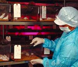
The modified adenovirus, called hTERT-Ad (human telomerase reverse transcriptase promoter regulated adenovirus), targets malignant glioma cells in mice using a process called autophagy, which is best described as self-cannibalism. Senior study author Seiji Kondo, said that after autophagy there was a reduction in tumor size and a marked survival rate among the mice.
The team found that those mice treated with three injections of hTERT-Ad lived an average of 53 days, while those receiving the control adenoviruses lived an average of 29 days. On average, the tumor size in the hTERT-Ad group was 39 cubic millimeters, while those receiving the control adenoviruses had an average tumor volume of 200 cubic millimeters. Even more encouraging were 2 hTERT-Ad treated mice that survived for 60 days without any sign of brain tumors.
This is great news, because other types of cancer are also telomerase-positive, and researchers have already shown in the lab that the virus can kill human prostate and human cervical cancer cells without affecting healthy tissue. In addition to these results, the team was able to elucidate on the processes by which such conditionally replicating adenoviruses (CRAs) infect and kill cancer cells. They explain that autophagy is a protective cannibalistic mechanism that the cell uses to eat or destroy some of itself when nutrients are scarce, or, for component recycling. The process consists of a double membrane enveloping an area to be consumed, and then digesting the membrane’s contents (burp!). Kondo showed that after infecting glioma cells, hTERT-Ad induced autophagy by deactivating a molecular pathway (the mammalian rapamycin (mTOR) pathway) that prevents cellular self-cannibalization.
The team was sure that autophagy, and not the commonly known apoptosis, had occurred after examining the dead cancer cells, which showed virus remnants in the cell nucleus and autophagy vacuoles. The damage caused by the two processes is quite different. Apoptosis damages the cell nucleus and DNA without damaging cellular organelles, while autophagy destroys most of the organelles with little damage inflicted upon the nucleus. ”We believe that autophagy, but not apoptosis, mediates the principal anti-tumor effect of conditionally replicating adenoviruses,” Kondo said.
Until now, apoptosis has been the main focus of attention in regard to programmed cell death, but Kondo says that apoptosis and autophagy should be viewed as version types 1 and 2. Kondo adds that cell death is a relatively new field of cancer research, and that to improve therapeutics it is vital to have a greater understanding of the molecules that regulate autophagy. The research team is following up its recent findings by improving the efficiency with which cancer cells can be infected with the virus.


Comments are closed.