In new research that will profoundly impact our understanding of biological processes, Stanford University scientists have discovered that the way cells “talk” to each other is far more complicated and nuanced than previously thought.
“Think of cells as musicians in a jazz band,” said the study’s author Markus Covert, whose lab studies complex genetic systems. “One little trumpet starts to play, and the cells go off on their own riffs. One plays off of the other.”
Up to now, most of the scientific information gathered on cell signaling has been obtained from populations of cells using bulk assays. The new study, published in Nature, shows that scientists have been misled by the results of the cell-population-based studies. “Population studies have not revealed the intricate network of information one observes at the single cell level,” explains Covert. “What we see is that differences between cells matter. Even the nuances can play a role.”
Cell signaling governs basic cellular activities in the human body. The ability of cells to correctly respond to their environments is the basis of all development, tissue repair and immunity. A better understanding of how cells talk to each other could lead to new insights into how larger biological systems operate, and possibly lead to cures for such diseases as cancer, diabetes and autoimmune disorders.
To study individual cell reactions during the cell-signaling process, Covert’s lab joined forces with Stephen Quake, a bioengineer at Stanford. Quake invented the biological equivalent of the integrated circuit – the microfluidic chip – which enables the study of single cells. In this study, Quake and Covert put it to use to investigate inflammatory cell signaling. “This study is a beautiful biological application of microfluidic cell culture and really illustrates the power of the technology,” Quake said.
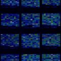
In the lab, the scientists put mouse fibroblast cells onto the chip and let them grow in an environmental chamber, which is mounted on an inverted microscope. The entire system, which fits on a small desktop, is computerized and provides long-term monitoring of the individual cell’s response to a signal by taking pictures every few minutes. For this study, Covert, Quake and their colleagues stimulated the cells with various concentrations of a protein that typically elicits the immune system’s response to infection or cancer.
“What we found is that some cells receive the signal and activate, and some don’t,” said co-researcher Savas Tay. In the images, the scientists could see that the cells responded in various ways, with different timing and number of oscillations, yet their primary response, in many respects, was equal. “Previously, we used to see the cell as a messy blob of biological material, yet there is great engineering down there,” explained Tay. “We needed to use mathematical modeling to understand what is going on.”
“The cells were doing totally different things and we’ve been totally missing it,” Covert added.
Related:
Geometry influences stem cell differentiation
Microbe Colonies Show Sophisticated Learning Behaviors


Comments are closed.